In the upcoming future, my teammates and I will be working with the Scanning Electron Microscope or SEM. We are especially excited about this because we can take a sample of biochar and be able to analyze certain properties under extreme magnification. The beauty of the SEM is that it has the capacity to magnify upwards to 500,000x. Our lab technician mentioned that once you get beyond a certain point, the picture tends to get a little grainy and sometimes difficult to handle. I only really plan on going as far as 200,000x magnification but I’m certain I’ll play around with more and if any image presents itself as being worthy of a photo, rest assured I will snap it! SEMs are pretty amazing in the way that they are able to examine a sample.
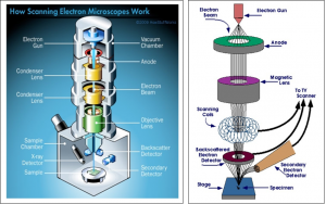
Basically, the scanning electron microscope fires a beam of electrons through a filament down at the sample you have staged. The electrons are kept in alignment through a series of alignment coils and condenser lenses which allow for a more concentrated beam making contact with the sample. As the electrons make contact with the sample, they begin to replace electrons from the atoms of the sample, itself. As these electrons from the sample are “deflected,” an electron detector collects them and begins to translate that as an image displayed on a computer screen. Simple enough in a sense.
Now in the regard to biochar we can begin to see a lot. For instance, different feedstocks will produce different types of biochar. Some of the major differences in the biochar are size, structure, shape, porosity, and mineral composition. Using the SEM we will be able to analyze these differences and actually be able to see the physical diversity of our samples. Knowing that we would be working with the SEM, I made sure to order a book that had a lot of relevance – Biochar: A Guide to Analytical Methods. This book broke down a lot of the various methods to examine the properties of biochar. Not only that, but it began with proper sample collection and storage depending on which tests you were planning to run. In this book they have a chapter dedicated solely to the SEM and analyzing biochar. Essentially, it mentioned that I may need to coat my sample with a conductive metal, also known as sputtering so that there are electrons available to be reflected and therefore a generated image. The metal is typically palladium, gold, chromium or platinum. The book also mentions that using a backscatter electron detector, we may also be able to visually see the different mineral phases of each biochar sample.