In this post I will cover the internal organs and anatomy of insects. The function of these anatomical pieces will come in my next post, and this serves more as an introduction to the various parts and their titles.
Endoskeleton: While the exoskeleton is the principal structure of the insect body, the endoskeleton still is important. It serves to strengthen the body will, and is another attachment point for muscles. A group of apophyses (spinelike or armlike processes) connects the endoskeleton to the head, forming the tentorium. This structure is usually H, X, or π shaped.
Muscular system: Insects can have between several hundred muscles and a few thousand. These are composed of striated muscle cells (marked by stripes, used for skeletal muscles in humans). These muscles are attached to the body wall and move the different parts of the insect. They typically connect to the body via tonofibrillae, which are fine connective fibers.
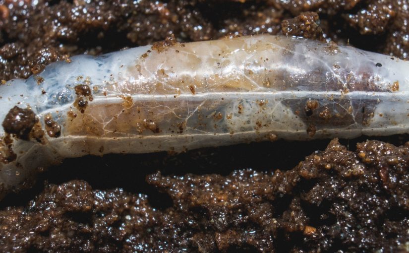
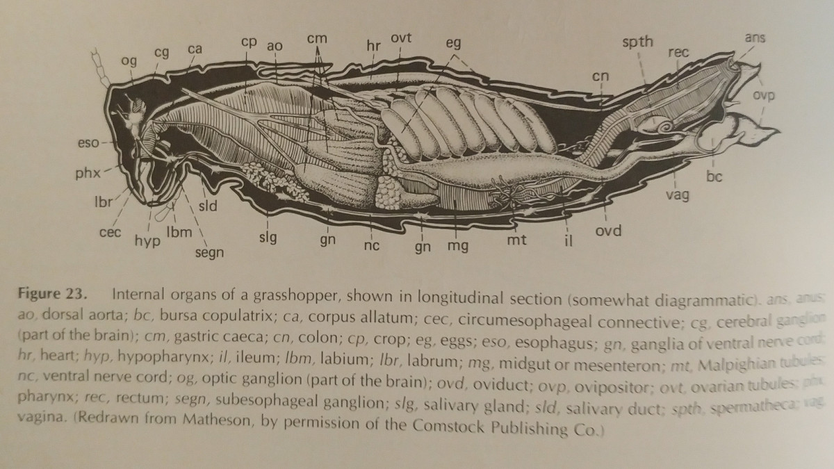
Digestive System: It’s important to note that different types of diets require different systems, and a large amount of variation is present. One of the main parts of the digestive system is the alimentary canal, a tube that runs from the mouth to the anus. This is visibly in the featured image of this post; it is the long, central tube in the larvae. There are three main regions of the digestive system: the stomodaeum (foregut), mesenteron (midgut), and the proctodaeum (hindgut). Various valves and sphincters regulate the travel of food from each section.
- Stomodaeum (foregut): Usually differentiated into a pharnyx (inside the mouth), a esophagus (a tube extending posteriorly from pharnyx), a crop (enlargement of the end of the esophagus), and the proventriculus (often bears teeth). At the end, the stomodaeal valve regulates transfer of material to the midgut. The foregut is lined by a small layer of cuticle known as the intima, which often has short hairs. Another part of the digestive system is the labial glands. Most insects typically have a pair underneath the anterior part of the alimentary canal. These are often called salivary glands, but they don’t always secrete saliva; Lepidoptera and Hymenoptera larvae use these glands to secrete silk.
- Mesenteron (midgut): Typically an elongate sac of uniform diameter, but sometimes is differentiated into two parts. It lacks a cuticle, and doesn’t have mucus to lubricate food. The lack of mucus also means no protection for epithelial cells, which are typically found between the intima and the longitudinal muscles above it. Therefore, these epithelial cells secrete a thin membrane, known as the peritrophic membrane. It is permeable, allowing the exchange of digestive enzymes and digested foods that are ready for absorption.
- Proctodaeum (hindgut): Extends from pyloric valve, which is between it and the midgut. It is supported posteriorly by muscles that extend to the abdomen wall, and is usually differentiated into two regions, the anterior intestine and rectum. The malphigian tubules are also located here, and they are how the insect excretes waste.
The final part of the digestion system is the filter chamber. Most Homoptera (aphids, etc) posses this. It is a modification of the alimentary canal, where the anterior part of the hindgut and the anterior part of the midgut are connected. These are usually quite distant. The midgut is differentiated into an anterior enlargement behind the stomodaeal valve, which is enclosed in the filter chamber, a croplike sac behind that, and then a long tubular section that turns to reenter the filter chamber. This is used to extract water from the food entering the midgut.
Circulatory System: Compared to a vertebrae, the circulatory system is rather open. The only blood vessel is a tube under the alimentary canal. The posterior part of this is the heart. It’s divided into many chambers by valves, with each chamber possessing an ostia (lateral opening), which is where blood enters. The anterior piece is slender, and is called the dorsal aorta. Outside of this vessel, blood simply circulates throughout the body cavity. Pairs of sheetlike muscle bands extend from the bottom of the heart to the lateral portions of the terga, which is known as the dorsal diaphragm. This serves to separate the heart from the rest of the body cavity.
Blood of insects is typically clear, sometimes green or yellowish. Cells called hemocytes are suspended inside. The blood typically makes up 5-40% of the body weight of an insect, and it bathes the organs and the tissues of the body. It is notable that the blood does little to transport oxygen as it does in our bodies.
Trachael System: Responsible for the intake and distribution of oxygen, as well as the removal of Carbon Dioxide. This is accomplished through many tubes, known as tracheae. These branch throughout the body, breaking off into finer segments called tracheoles, >1 micron thick intracellular tubes, which then permeate the tissues with oxygen. There are two types of systems, open and closed. In open systems, present in most insects, the spiracles can open. Closed systems have their spiracles permanently closed, and instead have a network of tracheae under their shell.
Excretory System: Consists of a group of tubes, known as the Malphigian tubules, which occur at the anterior end of the hindgut. The number various, however 1-200 of these tubes may be present. Waste is taken from the blood to the tubules, and then passed through the hindgut and anus. Occasionally, glands in the rectum may absorb water and salt.
Reproductive System: Insects primarily reproduce through sexual means, and usually posses distinct sexes. Parthogenesis, or unfertilized asexual reproduction, does occur in some species, such as eusocial bees or aphids, and some species even have no known males. Gonads are present in the abdomen, with ducts opening to the posterior end of it.
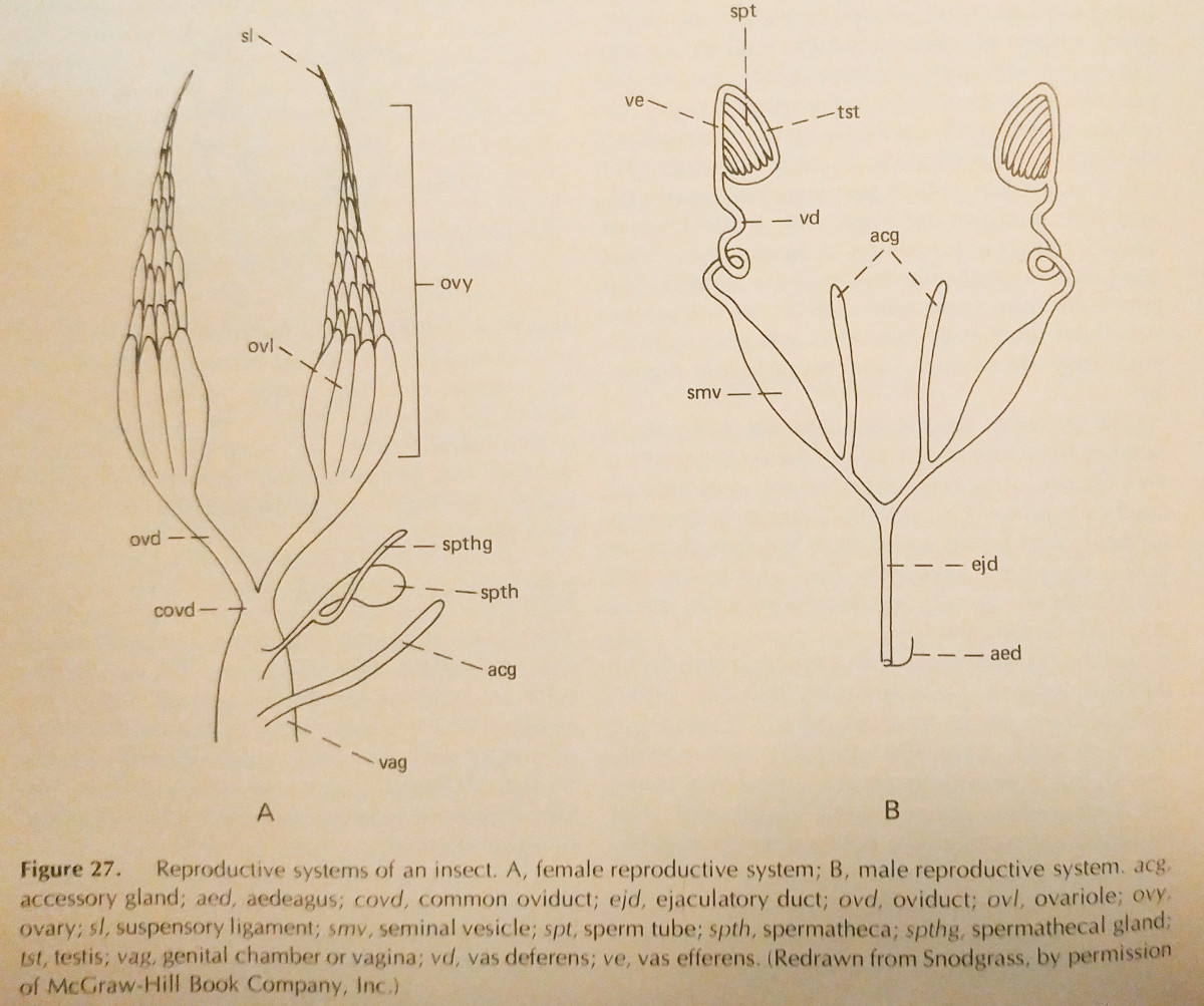
- Female Reproductive System: Females posses a pair of ovaries, a system of ducts for transporting the egg, and associated structures. Directly below the ovaries are a group of ovarioles, which lead to the oviduct posteriorly, uniting in a suspensory ligament that attaches to the body wall. Usually four to eight ovarioles are present, but 1-200 are possible. Often a spermatheca is present, a small sac in which sperm is stored. Adhesive glands for securing eggs may also be present.
- Male Reproductive System: Primarily, there is a pair of testes. These are attached to sperm tubes, which then attach to the Vas Deferens by stalklike vas efferens. There are many variations on this, as well as associated accessory glands.
The Fat Body is an aggregation of cells in the body cavity used for intermediate metabolism and food storage. It is usually best developed in the later instars or larval stages, and may be depleted after metamorphosis occurs.
Nervous System: The brain connects to a sub-esophageal ganglion via two commisures. In the previous post I mentioned that there were two lobes of the brain, however Borror, De Long, and Triplehorn, an updated reference from Snodgrass, mentions three. They are the protocerebrum (vision), deutocerebrum (antennal), and tritocerebrum (innervates labrum and foregut).
Sense Organs are located mainly within the body wall. Most are microscopic in size and are excited by a specific stimulus.
Chemical Senses are taste and smell. These structures are variable, but usually consist of a group of sensory nerve cells whose distal processes form a bundle that extends to the surface of the body. Taste is typically a product of the mouthparts, however some insects have taste organs on their antennae, or even the tarsi of their feet (Lepidoptera and Diptera).
Mechanical Senses are that of touch, pressure, or vibration. There are three types of structures responsible for this. The first type are hair sensillum, the simplest tactile receptor. They are simply a seta (hair) with a nerve cell. Next are the campaniform sensillum, which lack seta, and instead have the nerve ending in a domelike area beneath the cuticle. The Scolopophorous organs are more complex than the prior two. They are bundles of sensory cells attached to the body wall, and are widely distributed around it. They are sensitive to the movements of the insects body. Some examples of these include subgenual organs, Johnston’s organs, and tympanal organs.
Auditory Organs: Hair sensilla and tympanal organs are utilized to detect airborne sounds, however which hairs serve this function is not always clear. Tympanal organs are Scolopophorous organs with the sensory cells attached to tympanic membranes. These membranes contain air on both sides and are quite thin.
Vision Organs: These structures are sensitive to light. As I’ve noted in prior posts, these structures are the ocelli and compound eyes. Ocelli are composed of a single corneal lens that is elevated or domelike. There are two layers behind this lens, the corneagenous cells and the retina. Compound eyes are composed of many (up to several thousands) of individual ommatidia. They’re elongate groups of cells that are capped by a hexagonal cornea lens.
These are many of the organs and structures you may find within an insect. Of course, this list is nowhere near exhaustive, and I have even left out many terms that seemed too obscure. Now that the structures are outline, we can begin to discuss exactly what functions they serve for the insect!