
Glaucus atlanticus, blue sea slug, is a small, blue sea slug, pelagic nudibranch. G. atlanticus has a single row of a homodont tooth along a thin radular ribbon. G. atlanticus preys on other larger pelagic organisms such as the venomous Physalia physalis (Portugese man o’ war), the violet snail Janthina janthina, and by-the-wind sailor Velella velella.
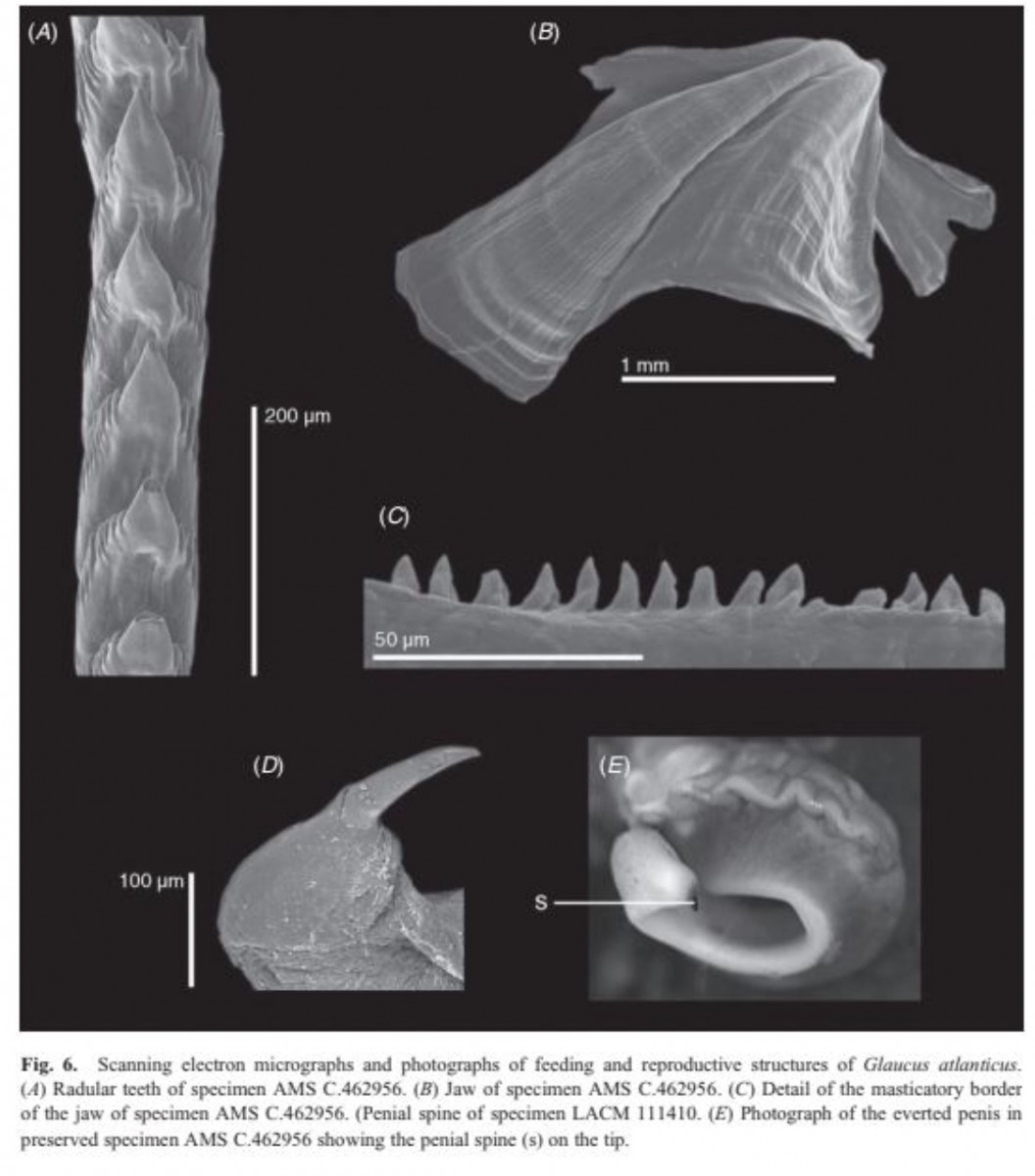
Churchill, C. K. C., Valdés, Á., & Ó Foighil, D. (2014). Molecular and morphological systematics of neustonic nudibranchs (Mollusca : Gastropoda : Glaucidae : Glaucus), with descriptions of three new cryptic species. Invertebrate Systematics, 28(2), 174. doi:10.1071/is13038
=====
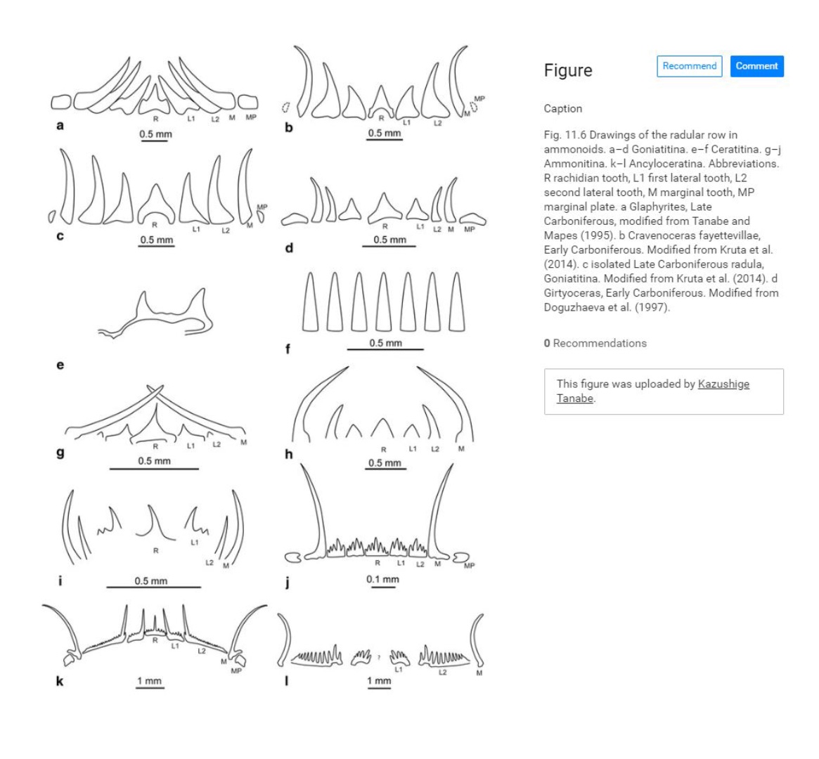
Chapter 11: Ammonoid Radula
Book: Ammonoid
By: Isabelle Kruta, Neil H. Landman and Kazushige Tanabe
=====
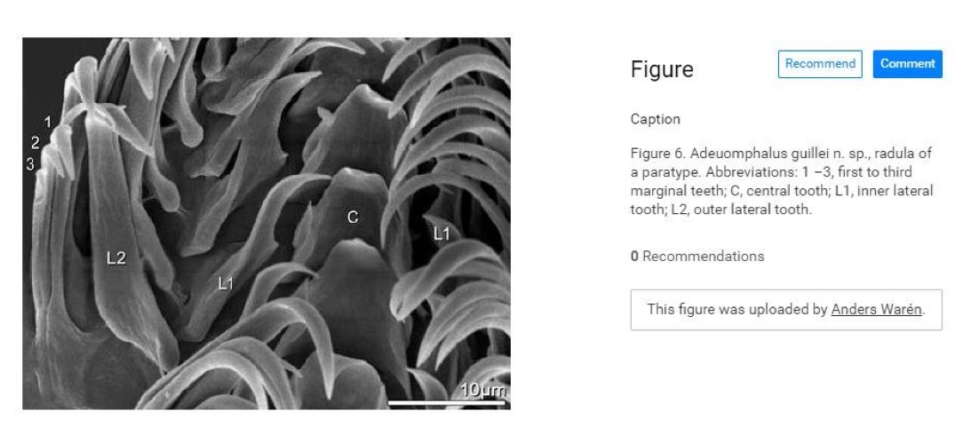
Kano, Y., Chikyu, E., Waren, A., (2009). Morphological, ecological and molecular characterization of the enigmatic planispiral snail genus Adeuomphalus (Vetigastropoda: Seguenzioidea). Journal of Molluscan Studies 75(4): 397-418.
=====
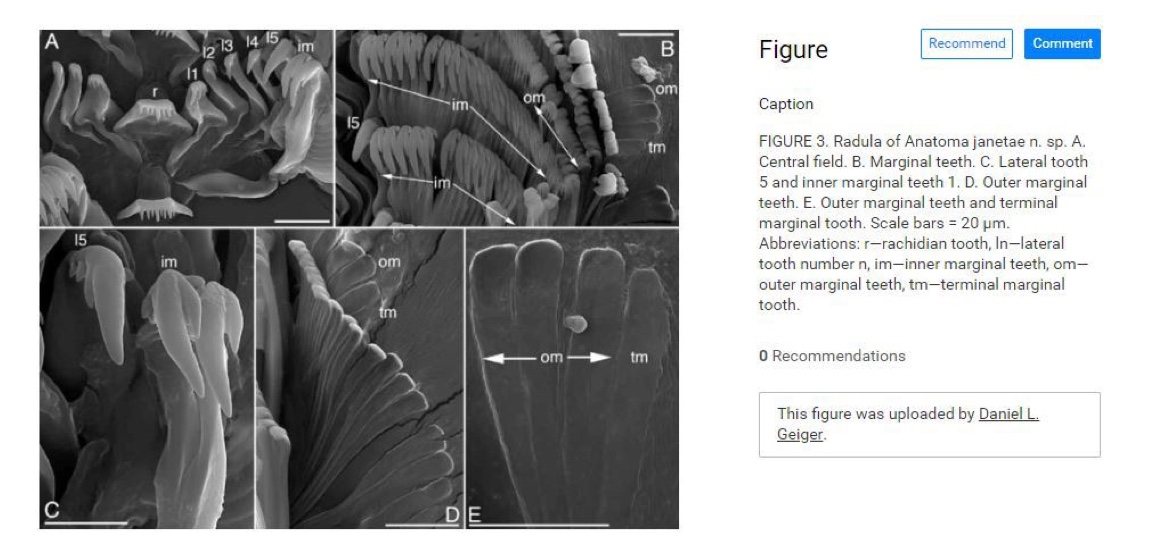
Geiger, Daniel L.. (2006). A new blind Anatoma species from the bathyal of the northeastern Pacific (Vetigastropoda: Anatomidae). Molluscan Research. 26. 108-112.
=====
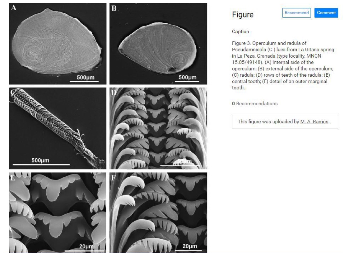
Delicado, Diana & Machordom, Annie & Ramos, M. (2012). Underestimated diversity of hydrobiid snails. The case of Pseudamnicola (Corrosella) (Mollusca: Caenogastropoda: Hydrobiidae). Journal of Natural History. 46. 25-89. 10.1080/00222933.2011.623358.
=====
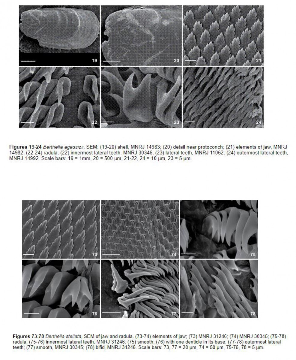
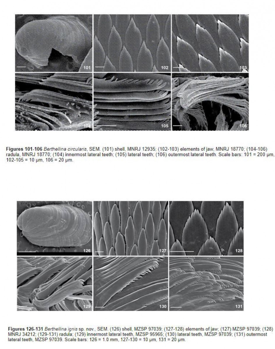
Alvim, J., Pimenta A.D., (2015). Taxonomic review of Berthella and Berthellina (Gastropoda: Pleurobranchoidea) from Brazil, with description of two new species. Zoologia (Curitiba) 32(6). http://dx.doi.org/10.1590/s1984-46702015000600010
=====
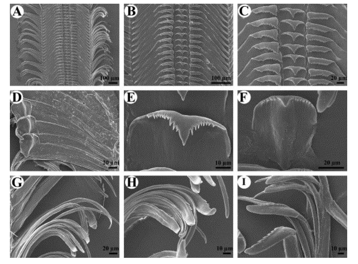
Fig. 3. Radula of T. horei. (A) Section of radular ribbon near anterior one-third (ZMB 220.095); note long extensions on lateral teeth. (B) Lateral and rachidian teeth (ZMB 220.095). (C) Detail of cusps of rachidian and lateral teeth (ZMB 220.095). (D) Detail of lateral teeth of second specimen at slightly different angle (MCZ 30.1576); note variation in laterals between specimens, and prominent pointed projection at inner edge of tooth base. (E) Detail of rachidian tooth (ZMB 220.095). (F) Detail of rachidian tooth of second specimen (MCZ 30.1576); note v-shaped base of rachidian, and variation between individuals in number of denticles. (G) Detail of marginal teeth (ZMB 220.095); note unequal numbers of fine denticles on inner and outer marginal teeth; also note rounded tip of inner tooth and pointed tip of outer tooth. (H) Detail of inner and outer marginal teeth (ZMB 220.095). (I) Detail of inner and outer marginal teeth of second specimen (MCZ 30.1576); note variation in number of denticles between individuals.
=====
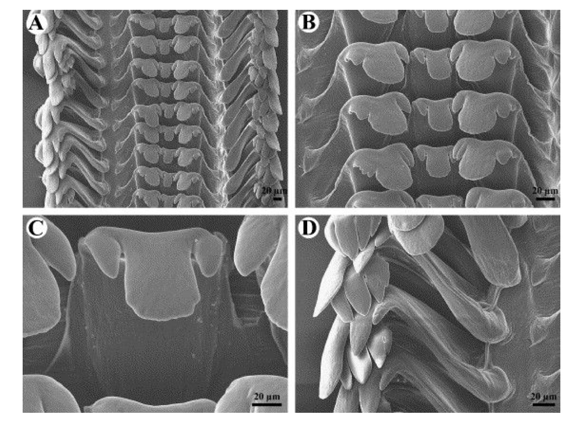
Fig. 12. Radula of Lavigeria sp. A (MRAC 621282–288). (A) Section of radular ribbon near anterior one-third. (B) Rachidian and lateral teeth. (C) Detail of rachidian tooth. (D) Marginal teeth; note unequal numbers of cusps on inner and outer teeth.
Strong E.E., Glaubrecht M., (2007). The morphology and independent origin of ovoviviparity in Tiphobia and Lavigeria (Caenogastropoda: Cerithioidea: Pauludomidae) from Lake Tanganyaki. Organisms Diversity & Evolution. 7(2):81-105.
======